Clinical Spotlight: Enhancing Vision and Comfort with a Recalibrated Scleral Lens
The Diagnostic Challenge
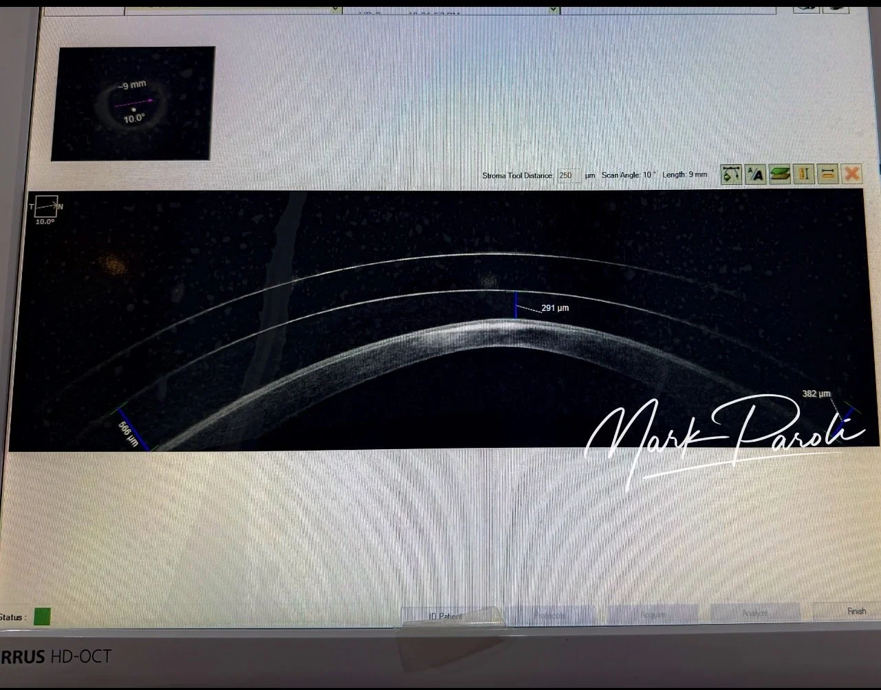
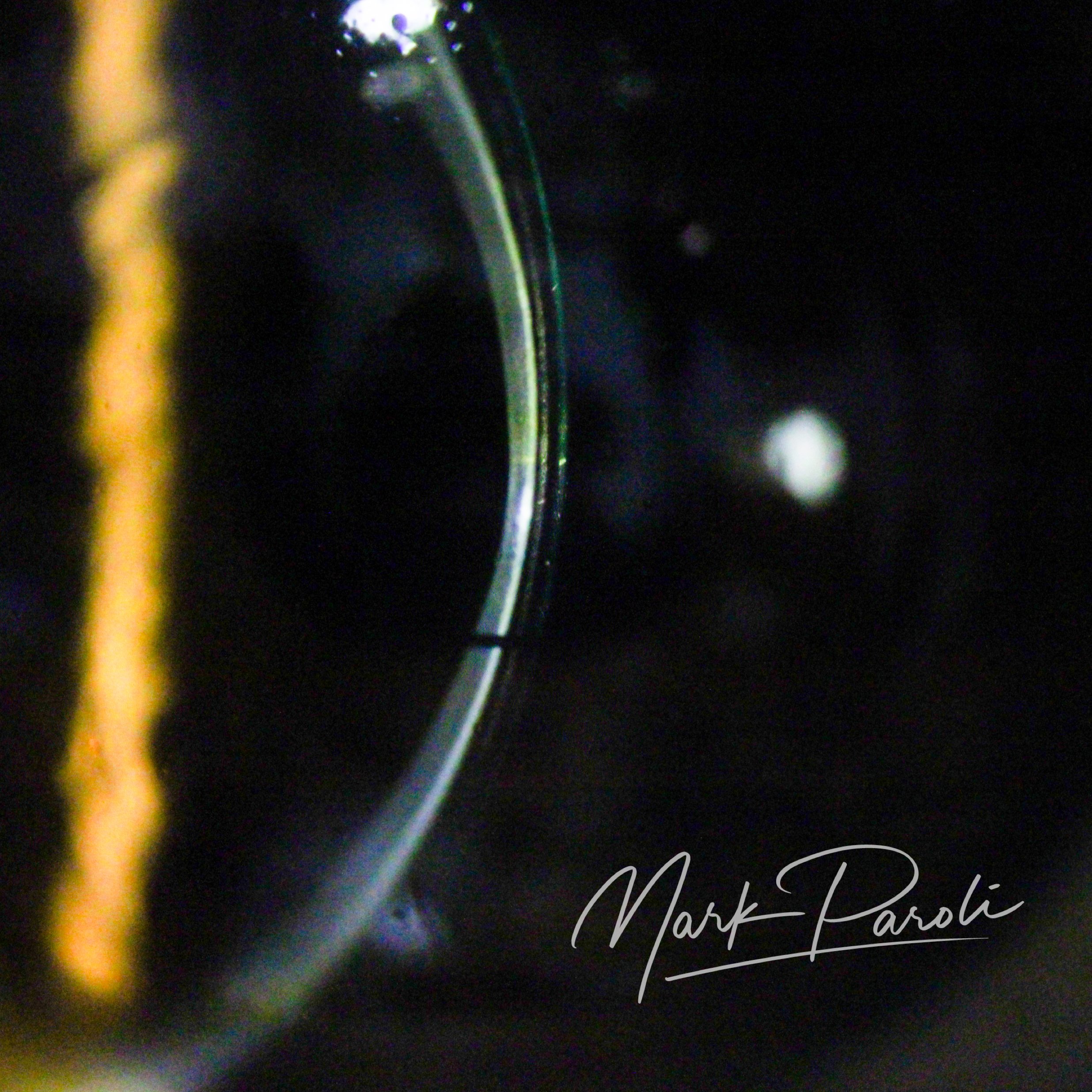
Anterior segment OCT scanning provides crucial insights when assessing and troubleshooting contact lens fits. A recent evaluation of a patient's previous scleral lens utilizing this technology revealed a central posterior lens tear and a fluid reservoir thickness of approximately 290 microns.
Precision Adjustments for Corneal Health
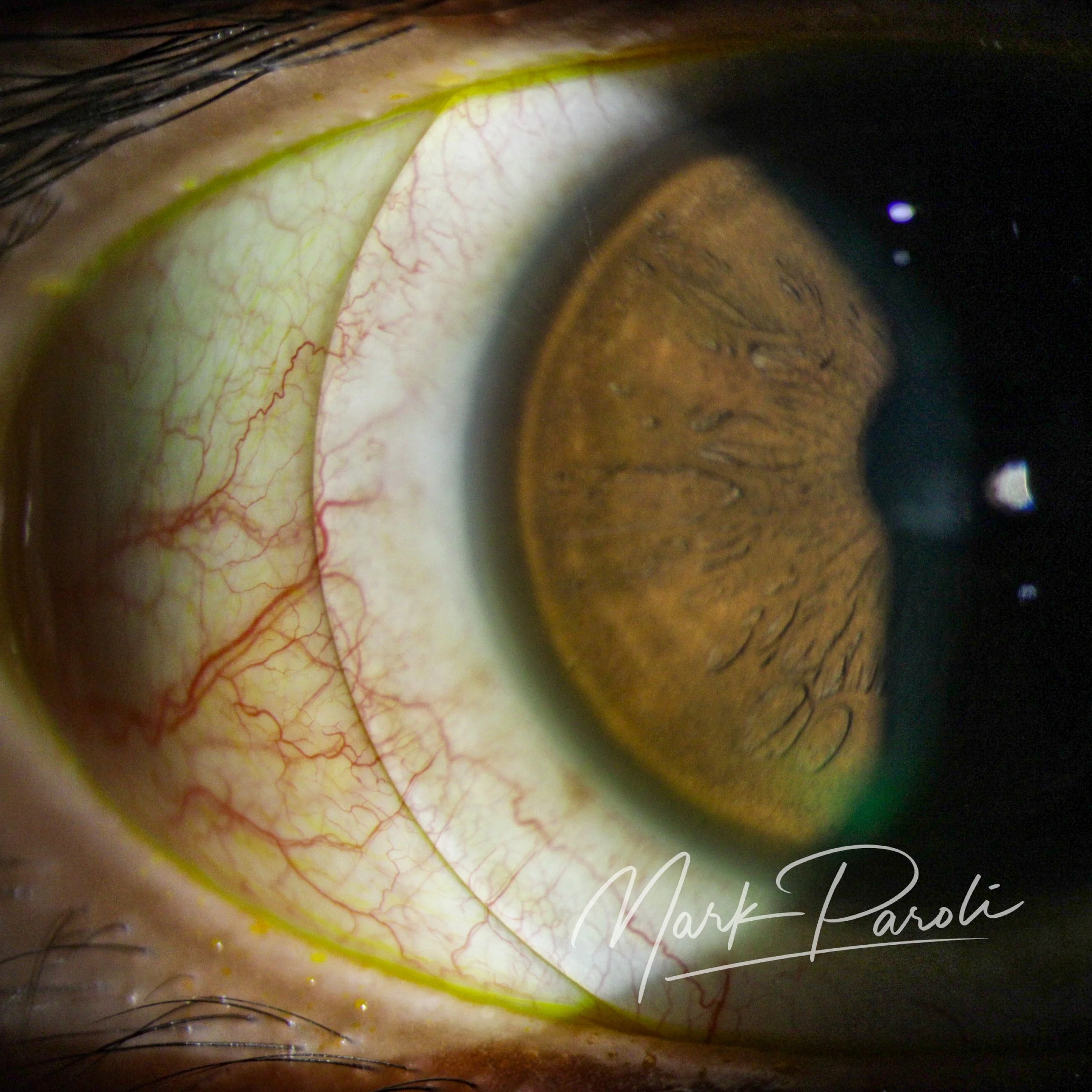
To enhance oxygen transmissibility and optimize the physiological environment of the cornea, the new lens required careful recalibration. The sagittal height was actively reduced, initially targeting a clearance of 150 microns. The final settled measurement stabilized at approximately 200 microns—striking an effective balance between corneal oxygenation and an appropriate vault.
Refractive Optimization
Once the physical fit was secured, visual acuity was refined through precise over-refraction. After confirming there was no significant rotation of the scleral lens on the eye, specific astigmatism correction was integrated directly into the lens optics.
The Patient Outcome
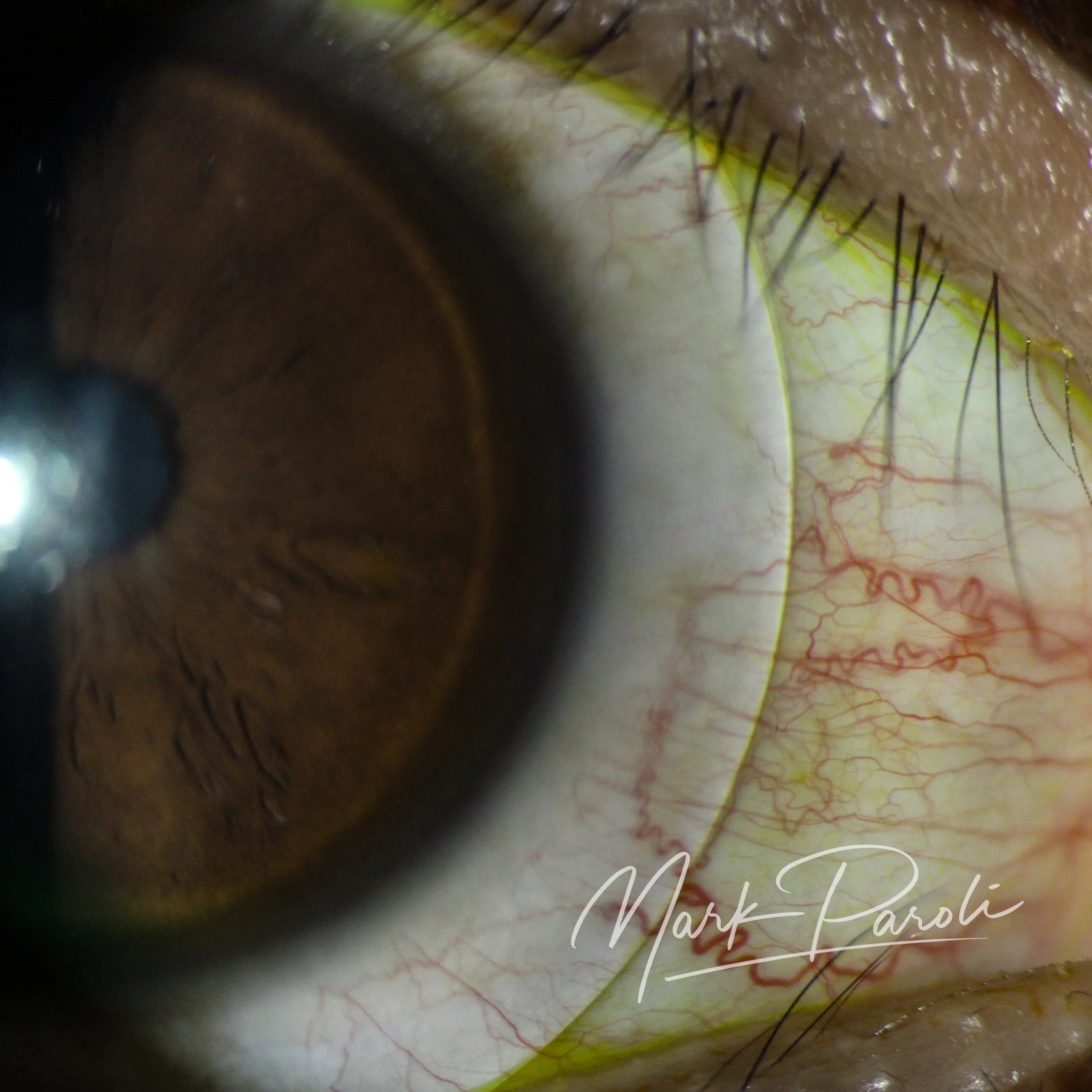
The combination of advanced imaging, targeted sagittal height reduction, and precise refractive correction resulted in a highly satisfied patient. The recalibrated fit delivered a significant improvement in visual clarity compared to their initial lens design.